Appointment Timing - 9am to 6pm

Liver cancer
Should This Giant Liver Tumor Be Resected?
34-year-old gentleman visited LPC, Mumbai with vague abdominal complaints and an USG & CT scan showing a large liver mass of unknown nature. He only had complaints of dyspeptic nature for few months. He did not have abdominal pain, vomiting, anorexia, jaundice, and fever, weight loss or GI bleed. There was no history of blood transfusion, previous liver disease or surgery. He did not have any co morbid factors like diabetes, hypertension, thyroid disorder, IHD, BA, TB, cirrhosis, kidney disease. He had never had alcohol and never smoked. General examination did not show any significant finding (pallor, cyanosis, jaundice, lymph nodes). Abdominal examination revealed a large palpable lump in the right hypochondrium, probably a mass arising from liver.
Since the imaging studies (USG & CT scan) he was carrying with him, were suboptimal i.e. not performed as per recommended international protocol for a liver tumor (triple phase liver protocol) and were also inconclusive it was decided to repeat them. Work up was started with blood investigation, which included CBC, LFT with PT/INR, tumor markers (AFP, CA19.9, CEA, Chromogranin A), HBsAg & anti HCV. All blood investigations including tumor markers were normal.
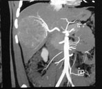
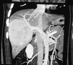
A triple phase IV contrast CT scan of abdomen& pelvis was performed. It showed a >10cm size liver tumor involving right lobe of liver. The tumor showed vascular enhancement in the arterial phase (hyperdense with surrounding liver parenchyma) with central necrosis. (PIC 1) In later / portal & delayed phase it became further hyperdense compared to surrounding liver parenchyma i.e. enhanced more. (PIC 2) These features were atypical for one particular tumor pathology and the differential diagnosis was a giant hemangioma (benign) vs Hepatocellular carcinoma (HCC—cancer) vs hepatic adenoma (potentially malignant) vs Focal nodular hyperplasia (benign). Since diagnosis could not be established a triple phase MRI was performed, which again gave similar possibilities.

PIC 1 - Arterial phase showing large tumor with vascular enhancement

PIC 2 - Portal phase showing further enhancement

PIC 3 - Angiography showing a highly vascularised tumor
Since a differentiation between benign and malignant was not possible a USG guided biopsy was done. Howeverbiopsy too was inconclusive. It showed mainly necrotic tissue. At places it showed atypical calls.As it was not possible to rule out a malignancy, a decision was taken to perform a hepatectomy. CT volumetry of liver showed that remnant liver volume after resection would be adequate (>35%). The lesion was very vascular. Hence a conventional angiography and tumor embolization was done one day prior to surgery with an aim to reduce intraoperative blood loss. (PIC 3).
Next day patient was taken up for a modified extended right hepatectomy. Exploratory laparotomy through a Mercedes Benz (modified bilateral sub costal) incision revealed a large vascular liver tumor (PIC 4). A modified extended right hepatectomy (right hepatectomy with a small area of left lobe i.e. liver segments 5,6,7,8 & 4b) was done with the tumor (PIC 5, 6, 7). Patient recovered uneventfully and was discharged after 8 days. Final histopathology & immunohistochemistry confirmed a well-differentiated hepatocellular carcinoma justifying our decision to go ahead with the surgery.

PIC 4 - Intraoperative picture showing a large tumor in right lobe of liver

PIC 5 - Intraoperative picture showing remnant liver after modified extended right hepatectomy

PIC 6 - Uncut specimen with the tumor

PIC 7 - Cut specimen with the tumor showing areas of necrosis and hemorhhage
Important Points In The Treatment Of Large Liver Tumor
- Giant tumors are fairly common in liver.
- Giant tumor has multiple definitions; however size larger than 10cms is always accepted as giant. Size greater than 5cms is also sometimes considered as giant in some centers.
- They often present late.
- It could be a solid or cystic tumor.
- Common differential diagnosis for solid tumor includes primary malignant liver tumors like hepatocellular cancer, intrahepatic cholangiocarcinoma; benign liver tumors like hemangioma, FNH, adenoma, gall bladder cancer with liver invasion, metastasis from primary tumors like colorectal cancer, NET from GIT/pancreas/ bronchus, GIST. Uncommon lesions like sarcoma are also seen.
- Common differential diagnosis for large cystic tumors include a simple liver cyst, hydatid cyst, biliary IPMN, BMCN, (cystadenoma and cystadenocarcinoma)
- A diagnosis is obtained using multiple investigations like serum tumor markers (AFP, CEA, CA19-9, chromogranin A), imaging studies like triple phase IV contrast CT scan &/or MRI, PET-CT, special imaging studies like SRS scan, DOTA scan (DOTA-TATE, DOTA-PET) & tumor biopsy.
- Imaging features may not be classical and differential diagnosis is difficult. In this situation tests are repeated over a period to see if there are any changes that can suggest the diagnosis. In spite of all investigations, a perfect diagnosis may still remain elusive.
- Tumors often have large central necrosis &/or hemorrhage, which might give false negative or wrong biopsy report i.e. a malignancy will be missed. Hence repeat biopsy may be required.
- Large tumors are often very vascular and make a biopsy difficult.
- A histopathologist (HP) specialized in hepatobiliary (HB) pathology are vital in reporting. Slides and cellblocks may be sent for review to a dedicated HB HP if initial reporting is inconclusive.
- Immunohistochemistry (IHC) is always useful in getting a correct HP.
- Treatment options depend on the diagnosis.
- Benign tumors like giant hemangioma & FNH may be observed as long as patient is not symptomatic or do not have high-risk features for hemorrhage. However a potentially malignant tumor like adenoma always requires resection. Occasionally liver transplantation (LT) is suggested for extensive benign tumor, if a resection is not possible. This is especially true for hemangioma.
- Malignant tumors like HCC & cholangiocarcinoma have a different problem, as the background liver is often cirrhotic precluding a major liver resection. A major liver resection in cirrhotic patient is possible only if liver function is sufficient (Child A). LT is contraindicated in large HCC or cholangiocarcinoma.
- Large colorectal cancer or neuroendocrine tumor metastasis amenable to resection are advised resection in combination with chemotherapy. When resection is not possible immediately due to extensive disease or inadequate future liver remnant; an attempt is made to down stage the disease with chemotherapy and later a resection is planned.
- Whenever a liver resection is not possible due to inadequate size of remnant liver, an attempt is made to increase the size by embolizing relevant portal vein branch.
- Special techniques have been devised for resection of large liver tumors not amenable to standard techniques. These include techniques like ex-situ resection or ante-situm resection, in-situ resection under cold preservation, major vascular resection-reconstruction etcetera.
Large tumors are difficult to resects because
- Liver remnant may be small and inadequate for survival
- Vascular invasion when malignant
- Major vascular reconstruction required
- Major blood loss during surgery
- Longer operative times
- Prolonged recovery time
- Major expenses involved in the treatment
Conclusion
Large liver tumors are a special problem in liver surgery and need special surgical skills for treatment. All efforts should be done to establish the nature of tumor. A biopsy should be done only if absolutely necessary. Whenever in doubt it is better to resect the tumor. Surgery can be safely performed in most situations with a mortality risk of <5%. However morbidity is still high in liver surgery especially in patients with such giant tumors.
Disclaimer:
The views expressed in this article solely belong to the author. The information provided here is for educational and informational purposes only. This newsletter is for private circulation only.
For Consultation Available At:
Timing: Monday To Friday - 9am To 5pm & Saturday 9am To 2pm
(Consultation Only by Appointment)
Address: Lilavati Hospital, A-791, Bandra Reclamation Rd, Bandra West, Mumbai, Maharashtra 400050
For Appointment call: 09821046391
Timing: Saturday 9am To 10am
(Consultation Only by Appointment)
Address: 93, ACI Hospital, 95, August Kranti Rd, Kemps Corner, Cumballa Hill, Mumbai, Maharashtra 400036
For Appointment call: 09821046391
Consultation Only by Appointment
Address: Raheja Rugnalaya Marg, Mahim West, Mahim, Mumbai, Maharashtra 400016
